When your skin heals after an injury, surgery, or even a simple piercing, you expect the wound to close and fade into a faint line. But for millions of people, the healing process goes awry. Instead of a flat, pale scar, a firm, raised mass emerges—sometimes itchy, sometimes painful, and often deeply frustrating. If you’ve landed here, you’re likely trying to understand whether that stubborn raised scar on your body is a keloid or a hypertrophic scar, and more importantly, what you can do about it.
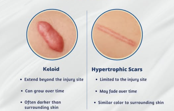
Here’s the core distinction: hypertrophic scars stay within the original wound boundaries and often flatten on their own over 12–24 months, while keloids extend beyond the original wound margins and rarely regress without intervention. Both result from abnormal wound healing and excessive collagen production, but they behave very differently over time. A hypertrophic scar may soften and fade; a keloid typically persists, may continue growing, and can cause relentless itching and tenderness for years. Keloids often cause more symptoms such as pain and itching compared to hypertrophic scars, which are primarily a cosmetic concern.
At HerbaScar™, we focus clinically on both keloids and hypertrophic scars because they share similar dermal remodeling challenges—dense, hardened collagen that surface creams simply cannot penetrate. However, we also recognize that each type demands different expectations and timelines. Keloid formation is influenced by genetic predisposition, dysregulation of cytokines like TGF-beta, inflammation, and mechanical forces after skin injury, leading to abnormal scar growth. Understanding which scar type you’re dealing with is the first critical step toward effective management and, ultimately, relief.
Quick Comparison Table: Keloid vs Hypertrophic Scar
A side-by-side comparison helps patients and clinicians quickly distinguish between these two types of abnormal scars. While both fall under the umbrella of pathologic scarring, their behavior, prognosis, and response to treatment differ substantially. The following table summarizes the key clinical differences.
Feature | Hypertrophic Scar | Keloid |
|---|---|---|
Spread beyond wound edges | No—stays within original wound boundaries | Yes—extends beyond original wound margins |
Onset timing | Weeks after injury (typically within 4–8 weeks) | Months to years after injury |
Tendency to regress | Often flattens and fades within 12–24 months | Rarely regresses; may continue enlarging |
Common locations | High-tension areas: shoulders, sternum, joints, surgical incisions | Earlobes, chest, upper arms, upper trunk, face |
Symptoms (itch/pain) | Mild to moderate itching, tenderness | Often intensely itchy, burning, painful |
Recurrence risk after excision | Lower (especially with adjuvant therapy) | High (45–100% without adjuvant treatment) |
Response to treatment | Generally good; many respond to silicone, pressure, or time | Often poor; requires multimodal, long-term approach |
It’s important to note that hypertrophic scars usually appear within weeks of wound closure and may improve significantly within 12–24 months, whereas keloids may emerge months or even years after the initial skin injury and rarely regress spontaneously. This table provides general guidance but is not a substitute for professional diagnosis—especially in individuals with darker skin tones, where visual appearance alone can be misleading.
Who Gets Keloids and Hypertrophic Scars?
Anyone can develop abnormal scars after a skin injury, but certain populations face dramatically higher risk. Understanding these risk factors helps identify who needs early preventive intervention and who should approach elective procedures with extra caution.
Keloids are significantly more common in people with darker Fitzpatrick skin types (III–VI), particularly those of African, Afro-Caribbean, Asian, and Hispanic descent. Epidemiological studies suggest that around 16% of Black individuals self-report keloids, making this a substantial public health concern in these communities. Hypertrophic scars, while also more prevalent in darker skin types, occur across all ethnicities—reported in up to 39–68% of surgical wounds and 33–91% of major burn injuries.
Hypertrophic scarring is especially common after deep burns, high-tension surgical incisions (such as those over the sternum or shoulders), and significant trauma. The mechanical forces pulling on the wound site during healing appear to drive excessive scar tissue formation regardless of genetic background.
Age plays a notable role as well. Peak incidence of both hypertrophic and keloid scars occurs between ages 11–30, when collagen synthesis is most active and skin tension tends to be highest. This explains why keloids occur most often in adolescents and young adults, who frequently develop keloids after ear piercings or acne on the chest and back. Keloids are less common than hypertrophic scars and are more likely to occur in younger populations aged 11 to 30 years.
Genetic predisposition is particularly strong for keloids. Family history significantly increases keloid risk, and specific HLA associations have been documented. Interestingly, Chinese populations appear to have higher keloid rates compared to some neighboring ethnic groups, suggesting complex genetic factors beyond simple melanin content.
Individuals with keloids may suffer from embarrassment and frustration due to their appearance and the failure of previous treatments.
Key risk factor summary:
- Darker skin types (Fitzpatrick III–VI)
- Age 11–30 years
- Family history of keloids
- High-tension body sites (shoulders, sternum, joints, earlobes)
- History of severe acne, burns, or major surgery
What Causes Keloids and Hypertrophic Scars? (Etiology & Risk Factors)
Both keloids and hypertrophic scars follow damage to the dermis—the deeper layer of skin beneath the surface epidermis. This damage can come from burns, piercings, surgery, acne, C-sections, tattoo removal, vaccinations, or even seemingly minor injuries. The key is that the wound must extend deep enough to disrupt the dermal architecture. Abnormal scar development often occurs during the healing wound phase, when the body’s repair mechanisms are most active—a process now increasingly referred to as wound repair regen, highlighting the importance of tissue regeneration and repair mechanisms in abnormal scar formation.
The exact biological “switch” that transforms normal wound repair into a keloid or hypertrophic scar remains incompletely understood. What we know is that it involves an overactive inflammatory response and overproduction of collagen by fibroblasts—the cells responsible for laying down new tissue. In predisposed individuals, this process spirals beyond what’s needed for wound closure, creating dense, raised scar tissue that serves no functional purpose. When evaluating hypertrophic scarring keloids, it’s important to note that hypertrophic scars remain confined to the original wound borders, are often red and raised but may regress over time, while keloids extend beyond the wound edges, are firmer, and display distinct histological features such as thicker collagen bundles and more disorganized tissue architecture. Keloid generation is driven by complex interactions between mechanical forces, chronic inflammation, and cellular signaling pathways that result in excessive collagen deposition. Hypertrophic scar formation, meanwhile, represents a dysregulated part of the normal healing process, where collagen production exceeds what is necessary for proper tissue repair.
Common triggers include:
- Ear piercings (especially cartilage piercings)—earlobe keloids are among the most common presentations
- Cesarean section incisions
- Sternotomy scars after heart surgery
- Shoulder and upper trunk trauma
- Acne on the chest and back
- Vaccinations and multiple needle punctures
- Thermal injuries (burns, laser treatments)
- Tattoos and tattoo removal procedures
- Healing wound sites subjected to excessive tension or trauma
Beyond local triggers, systemic factors influence risk. Chronic inflammation, hypertension, and hormonal changes (including pregnancy) appear to increase susceptibility. Areas of high mechanical tension or repeated stretching—such as joints, the chest, and shoulders—are particularly prone to forming hypertrophic scars and keloids.
One important distinction: hypertrophic scars almost always follow clear trauma or surgical procedure. Keloids, on the other hand, may appear after very minor injuries, folliculitis, or even seemingly “spontaneously” in highly predisposed individuals—sometimes emerging on skin where the patient cannot recall any injury at all.
Major modifiable risks to consider:
- Avoid unnecessary piercings in high-risk individuals, especially on earlobes, chest, and upper back
- Choose experienced surgeons who use careful closure techniques in high-tension areas
- Manage acne aggressively to prevent deep inflammatory lesions
- Consider alternatives to elective surgeries or cosmetic procedures if you have a history of keloids
To help prevent keloids and hypertrophic scars, it is important to minimize tension on healing wounds during the recovery process.
How Do Keloids and Hypertrophic Scars Form? (Pathophysiology)
Normal wound healing proceeds through three overlapping phases: inflammation (the body’s initial response to injury), proliferation (new tissue formation), and remodeling (maturation and organization of the new tissue). In healthy healing, these phases complete within months, leaving a flat, soft scar that blends with surrounding normal tissue.
In hypertrophic scarring and keloids, this process becomes dysregulated. The inflammatory phase extends beyond its normal duration, fibroblast activity becomes excessive, and collagen deposition in the dermis far exceeds what’s needed. Studies estimate that collagen production is approximately 3× normal in hypertrophic scars and can reach up to 20× normal in keloids—explaining why these scars feel so hard, dense, and elevated.
Several cytokines and growth factors drive this pathological process. Transforming growth factor beta (TGF-β1 and TGF-β2) levels are elevated, while TGF-β3—which normally helps limit scarring—is decreased. Pro-inflammatory markers like IL-6 and IL-8 remain elevated, and IL-10, which has anti-fibrotic properties, is altered. In simple terms, the signals telling the body to “keep building” overwhelm the signals saying “that’s enough.”
The structure of the collagen fibers themselves differs between these scar types. Hypertrophic scars contain more organized, parallel collagen fibers that generally align with skin tension lines—still abnormal, but somewhat orderly. Keloids, by contrast, feature thick, haphazard collagen bundles that disregard original wound margins entirely. Keloid collagen is often described as “hyalinized”—dense, eosinophilic, and arranged in whorled patterns that expand outward from the wound. Notably, keloids often lack hair follicles and other functioning adnexal glands, which helps distinguish them from benign or malignant tumors originating from hair follicle structures.
Histologically, hypertrophic scars have collagen arranged in a wavy, regular pattern, while keloids have collagen arranged in a haphazard pattern without a distinct structure.
This dense, disordered collagen architecture directly causes symptoms. Trapped nerve fibers within the thickened dermis contribute to the intense itching, burning, and sharp pain that many patients with keloids and hypertrophic scars report. The tissue is essentially “too full”—packed with excess collagen, blood vessels, and inflammatory cells that create constant irritation.
Key biological differences:
- Hypertrophic scars: organized collagen fibers, self-limiting growth, often resolve over 1–2 years
- Keloids: disorganized, hyalinized collagen bundles, progressive growth, rarely regress without treatment
- Both: elevated TGF-β, excessive fibroblast activity, increased collagen synthesis, trapped nerve fibers causing itch and pain
Keloids and hypertrophic scars can lead to prolonged pain and pruritus, causing emotional and psychological distress to patients.
For a comprehensive overview of keloid pathophysiology, treatments, and management strategies, see the literature on “keloids a review.”
What Do Keloids and Hypertrophic Scars Look and Feel Like? (Clinical Features)
Living with a raised scar is more than a cosmetic concern. The constant itching, tenderness, and tightness can affect sleep, clothing choices, intimacy, and self-confidence. Many patients describe feeling embarrassed to wear short sleeves, swimwear, or jewelry that draws attention to their scars. Understanding exactly what each scar type looks and feels like helps you identify what you’re dealing with—and communicates more effectively with healthcare providers.
Hypertrophic scars present as raised, firm lesions that are pink to red in color. Crucially, they stay within the boundaries of the original incision or injury—they don’t “spread” beyond where the skin was actually damaged. Hypertrophic scars tend to be contained within the injury site and may regress over time, often flattening and fading significantly over 12–24 months, especially in certain anatomical locations and under specific conditions. They may be itchy or tender, especially in the first year. Common locations include shoulders, the sternum, joints, and along surgical incisions. The scar tissue typically follows the line of the original trauma.
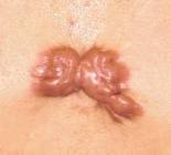
Keloids are also raised but often appear more bulbous or nodular. The defining feature is extension beyond the original wound borders—sometimes with irregular, “claw-like” projections that spread into surrounding normal tissue. Keloids tend to be shiny, smooth, and hairless, with colors ranging from pink to purple to brown depending on skin tone. Unlike hypertrophic scars, they rarely regress spontaneously and may continue to enlarge over months or years. Keloids can continue to develop for up to one year and do not spontaneously regress, leading to ongoing psychological distress. Earlobe keloids, upper trunk keloids, and those on the shoulders commonly occur.
The texture differences are notable. Hypertrophic scars feel firm but generally follow a linear pattern matching the original wound. Keloids may be lobulated (bumpy), nodular, or even pedunculated (attached by a stalk), especially on earlobes and the neck. Some keloids on the abdomen or chest become quite large and irregularly shaped.
Both scar types can cause pruritus (itching), burning sensations, and pain—but keloids are frequently reported as more intensely itchy and tender. Some patients describe sharp, shooting pains or constant throbbing. Functional impact is also significant: scars crossing joints or high-tension areas can restrict movement of shoulders, elbows, knees, or neck, contributing to scar contractures that limit daily activities.
Visual “red flags” suggesting a lesion is more likely a keloid:
- Growth clearly extends beyond the original wound edges
- Continued enlargement after 6+ months
- Irregular, spreading, or claw-like borders
- Intense, persistent itching despite treatment attempts
How Do Skin Type and Pigmentation Affect These Scars?
Melanin and baseline skin pigmentation significantly influence how scars appear and how they are perceived by both patients and clinicians. This matters for diagnosis, treatment planning, and setting realistic expectations.
Keloids are substantially more frequent in individuals with darker skin phototypes. On dark skin, keloids tend to appear less “red” and more hyperpigmented—often brown, violet, or dark purple rather than the pink-red typical in lighter skin. This can make early keloids harder to distinguish from post-inflammatory hyperpigmentation or other skin changes.
Post-inflammatory hyperpigmentation commonly surrounds or overlays both keloids and hypertrophic scars in darker skin, making the scar boundaries harder to define visually. A scar that looks small on the surface may actually extend deeper and wider than it appears. In very fair skin, early hypertrophic scars often appear bright pink or red; in darker skin types, they may blend more with surrounding tone while remaining distinctly firmer and raised to the touch.
This visual complexity underscores the importance of touch, patient history, and sometimes dermatoscopic or histologic examination in darker skin tones. Relying on color alone can lead to misdiagnosis, delayed treatment, or inappropriate reassurance that a lesion is “just a regular scar.”
Practical points for patients with darker skin:
- Higher risk of keloids with piercings, tattoos, and C-sections—consider avoiding elective procedures in high-risk areas
- Early preventive care (silicone sheets, compression therapy, or protective botanical extracts) is especially important
- Seek evaluation if a scar continues growing or becomes symptomatic
- Don’t assume a flat-looking hyperpigmented area isn’t problematic—firmness and expansion matter more than color
Diagnosis: Distinguishing Keloids from Hypertrophic Scars
Diagnosis of hypertrophic and keloid scars is primarily clinical, based on patient history and physical examination. Hypertrophic scars diagnosed in clinical practice rely on a detailed evaluation of the scar’s onset timing, growth pattern, borders relative to the original wound, and associated symptoms like itching or pain. When necessary, diagnosis may be confirmed with a skin biopsy to distinguish between scar types or rule out other conditions.
Timeline differences provide important clues. Hypertrophic scars typically appear within weeks of wound closure—usually 4–8 weeks—and remain confined to the wound site. Keloids may emerge months or even years after the initial skin trauma and characteristically extend beyond the original wound margins with irregular, sometimes “claw-like” extensions.
Key diagnostic features:
Characteristic | Hypertrophic Scar | Keloid |
|---|---|---|
Onset | Within weeks of injury | Months to years after injury |
Growth pattern | Confined to wound site | Extends beyond wound margins |
Natural history | Tends to regress over 12–24 months | Rarely regresses; may enlarge indefinitely |
Border appearance | Follows wound line | Irregular, spreading projections |
A skin biopsy may be required when diagnosis is uncertain or when there’s concern about other conditions that mimic scars—including certain tumors or inflammatory dermatoses. Histologically, hypertrophic scars show organized, wavy collagen fibers running parallel to the skin surface. Keloids display thick, whorled, disorganized collagen bundles, often with more mast cells and a hypocellular, hyalinized central zone.
When to see a dermatologist or plastic surgeon:
- Rapid or continuous growth of a raised scar
- Severe pain, burning, or itching that disrupts daily life
- Color change, ulceration, or bleeding from the scar
- Multiple spontaneous lesions appearing without clear trauma
- Any atypical appearance that doesn’t fit typical keloid or hypertrophic patterns
Differential Diagnosis: Conditions That Can Resemble Keloids or Hypertrophic Scars
Several benign and malignant skin lesions can mimic keloids or hypertrophic scars, making professional evaluation essential in atypical cases. A raised, firm nodule that doesn’t behave as expected deserves closer examination—not dismissal as “just a scar.”
Malignant mimickers include dermatofibrosarcoma protuberans (a slow-growing soft tissue sarcoma), keloidal basal cell carcinoma, and trichilemmal carcinoma. These conditions can present as firm, raised lesions that resemble keloids but require histology and immunohistochemistry for definitive diagnosis. Fortunately, they are rare—but missing them can have serious consequences.
Non-malignant look-alikes include sclerotic neurofibroma, cutaneous scleroderma plaques, thick hypertrophic lichen planus, and chronic infectious nodules or folliculitis scars. Some of these conditions require different treatments entirely and can worsen with inappropriate keloid therapies.
It’s worth emphasizing that true keloids and hypertrophic scars are benign. They do not themselves “turn into” skin cancer. However, any changing, ulcerating, rapidly growing, or highly atypical lesion should be biopsied to rule out malignancy—especially if it doesn’t respond as expected to standard scar management.
Warning signs that should prompt urgent evaluation:
- Ulceration, bleeding, or crusting over the lesion
- Very rapid growth (weeks rather than months)
- Pain out of proportion to typical keloid symptoms
- Lesions appearing on skin without any history of injury or trauma
Management and Treatment: Keloid vs Hypertrophic Scar
Let’s be honest: hypertrophic scars generally respond better to treatment and may resolve partially on their own; keloids are more stubborn, with higher recurrence rates even after surgical excision. According to evidence-based approaches and clinical studies published in dermatol surg, the scientific management of keloids and hypertrophic scars often requires a multidisciplinary strategy. Hypertrophic scar treatment and treating hypertrophic scars often involve a combination of therapies, including topical agents, intralesional injections, and laser procedures, to optimize outcomes and reduce recurrence. Surgical excision is sometimes considered, but excised keloids frequently require adjuvant therapies—such as corticosteroid injections or radiation—to minimize recurrence, as highlighted in plast reconstr surg, aesthetic plast surg, ann plast surg, and arch facial plast surg literature. This reality shapes treatment planning and patient expectations.
The goals of treatment include reducing scar height, softening texture, relieving itch and pain, restoring movement (for scars that limit function), and improving color to match surrounding skin as closely as possible. Complete “erasure” of a keloid is rarely achievable, but meaningful improvement in comfort and appearance is realistic for most patients.
Silicone Gel and Sheets
Silicone gel and silicone sheets remain first-line topical therapies for both scar types. They work by hydrating the scar and may help flatten early hypertrophic scars. However, they sit on the epidermis and often fail to penetrate the dense collagen matrix of established keloids.
Compression Therapy
Compression therapy applies constant pressure to the scar, reducing blood flow and collagen deposition. It’s most effective for hypertrophic scars on limbs or areas where occlusive dressings can be applied consistently.
Corticosteroid Injections
Corticosteroid injections (typically triamcinolone) reduce inflammation and collagen synthesis. They’re commonly used for both scar types but can cause skin atrophy, hypopigmentation, and pain. Multiple sessions are usually required.
5-Fluorouracil (5-FU) Injections
5-Fluorouracil (5-FU) injections inhibit fibroblast proliferation. Often combined with corticosteroids to reduce side effects while enhancing efficacy.
Cryotherapy
Cryotherapy freezes the scar tissue, causing cell death and volume reduction. Can be painful and may cause hypopigmentation, particularly in darker skin types.
Laser Therapy
Pulsed dye laser and other laser therapy options target blood vessels within the scar, reducing redness and potentially softening tissue. Multiple sessions are needed, and results vary.
Surgical Excision
Surgical excision removes the keloid or hypertrophic scar but carries significant risk of recurrence—often quoted at 45–100% for keloids when surgery is performed alone. It’s almost always combined with adjuvant therapy (steroid injections, radiation, or silicone) to reduce this risk.
Radiotherapy
Radiotherapy (superficial X-ray or electron beam) after excision significantly reduces keloid recurrence but raises long-term concerns about radiation exposure and is typically reserved for severe, recurrent cases.
Emerging Options
Emerging options include interferon injections, botulinum toxin (which may reduce skin tension), bleomycin tattooing, and TGF-β modulators—though most remain investigational or have limited availability.
Pros and cons summary:
Treatment | Pros | Cons |
|---|---|---|
Silicone sheets/gel | Non-invasive, low risk, easy to use | Limited penetration for dense keloids |
Corticosteroid injections | Reduces inflammation quickly | Painful, can cause atrophy/hypopigmentation |
Surgical excision | Immediate volume reduction | High recurrence without adjuvant therapy |
Laser therapy | Reduces redness, non-invasive | Multiple sessions, variable results, costly |
Compression therapy | Effective for accessible sites | Difficult to apply consistently on some areas |
Radiotherapy | Significantly reduces recurrence | Radiation exposure concerns, limited availability |
The critical point: monotherapy often fails in keloids. Combination approaches—such as surgical excision followed by corticosteroid injections and silicone application—are typically required to achieve lasting improvement and lower recurrence rates.
HerbaScar™ Approach to Keloids and Hypertrophic Scars
At HerbaScar™, we’ve spent over a century refining our understanding of deep tissue remodeling. Our clinical-grade botanical liquid extract system is rooted in a Traditional Chinese Medicine lineage dating back to 1907, now refined with modern 90-day cold-extraction technology that preserves maximum tissue-softening potency without harsh solvents or high heat.
Why Thick Creams Fail
Here’s the problem with most drugstore scar products: they’re thick creams or gels that sit on the epidermis—the skin’s surface layer. They cannot penetrate the hardened collagen matrix deep in the dermis where keloids and hypertrophic scars actually live. This is why so many people spend $20 on silicone sheets or onion-extract gels, apply them faithfully for months, and see minimal improvement. The product simply never reaches the dermal root of the problem.
The HerbaScar™ Liquid Extract Mechanism
HerbaScar™ uses a transdermal liquid delivery system that bypasses the epidermis to deliver active ingredients directly into the dermis. The key is Borneol—a natural penetrator that opens pathways through the skin barrier and provides immediate itch relief. Once deep penetration is achieved, other botanical actives go to work:
- Panax Notoginseng: The tissue breaker that helps soften dense, hyalinized collagen fibers
- Galla Chinensis: Supports natural shrinking and volume reduction of raised tissue
- Licorice Root Extract: Calms inflammation and brightens hyperpigmented surrounding skin
The 3-Stage Remodeling Cycle
We manage expectations honestly. Dermal remodeling doesn’t happen overnight—it’s a gradual process requiring consistency over weeks and months.
Stage 1: Penetration & Softening The liquid extract penetrates the hardened scar tissue. Patients typically notice reduction in itching and burning first, followed by gradual softening of the raised area.
Stage 2: Cellular Shedding As the dense collagen matrix breaks down, a controlled medicated scab may form. This represents cellular shedding—dead, thickened tissue detaching as healthier tissue emerges beneath.
Stage 3: Protection & Prevention Fresh skin is vulnerable. This stage focuses on protecting the newly revealed tissue and reducing the risk of abnormal regrowth.
Matching Products to Scar Types
Not every scar needs the same formula. Our product lineup addresses different scar presentations:
- HerbaScar™ Advanced Scar & Keloid Liquid ($199): The hero product for raised scars. Best for keloids, hypertrophic scars, piercing bumps, and dense surgical scars. Mechanism: breaks down excess collagen and stops itching.
- HerbaScar™ Chitosan Keloid Defense Gel: The post-treatment protector. Best for keloid prevention, post-scab protection, and fresh incisions. Uses marine-derived biological shield technology to prevent abnormal scar tissue from returning.
- HerbaScar™ Laser & Tattoo Trauma Defense: The thermal rescue formula. Best for post-tattoo removal and laser burns. Rapidly cools thermal damage, calms extreme inflammation, and prevents raised outlines from forming.
- HerbaScar™ Atrophic Scar & Pit Resurfacing Serum: The rebuilder. Best for depressed scars like pitted acne scars or ice pick scars. Note: this formula stimulates collagen synthesis to “fill in” the dermal bed—the opposite mechanism of the keloid liquid, which breaks collagen down.
Safety and Quality
HerbaScar™ products contain 0% steroids, 0% silicone, 0% parabens. They’re cruelty-free and entirely non-invasive. For patients who have already endured painful steroid injections, failed surgeries, or frustrating experiences with cheap drugstore products, we offer a 90-day empty-bottle money-back guarantee. The risk is entirely on us.

How to Choose the Right Treatment Path for Your Scar Type
Correct classification is critical before starting any treatment. Using a collagen-breaking formula on a depressed (atrophic) scar—or a collagen-building serum on a keloid—will be counterproductive at best and potentially harmful.
Decision-support guide
- If the scar is raised but stays within the original cut or burn and is less than 2 years old: Think “hypertrophic” and consider non-invasive remodeling first. These scars often respond well to consistent daily application of deep-penetrating liquid extracts, silicone, or pressure therapy.
- If the scar clearly extends beyond the original wound, is intensely itchy or painful, and keeps enlarging: Think “keloid” and prepare for a longer, layered treatment strategy. Combination approaches work best, and consistency over many months is essential.
- If the scar is sunken or pitted: This is an atrophic scar requiring collagen-building therapies (like HerbaScar™ Atrophic Scar & Pit Resurfacing Serum), not collagen-breaking ones.
When to Prioritize In-Clinic Procedures
Very large keloids, those causing functional limitations (restricting joint movement), or keloids on cosmetically critical areas (face, neck) may require surgical excision with adjuvant therapy. However, even after excision, a home-based liquid remodeling approach helps maintain results and prevent recurrence.
When Home-Based Remodeling Is Appropriate
Small to medium raised scars, early post-surgery scars, and earlobe keloids often respond well to consistent topical therapy—provided the formulation actually penetrates to the dermal level. This is where clinical-grade liquid extracts outperform surface creams.
The key principle: Consistency is more important than aggressiveness. Daily application for months typically outperforms sporadic, painful interventions done only once or twice.
Before consulting a dermatologist or HerbaScar™ specialist, prepare:
- Age of the scar (when did it first appear?)
- Cause of the original wound (surgery, piercing, acne, burn, trauma?)
- Previous treatments attempted and their results
- Current symptoms (itching severity, pain, growth pattern)
- Clear photographs showing scar size relative to a ruler or coin
Prevention: Reducing the Risk of Keloids and Hypertrophic Scars
Concrete Preventive Measures
- Careful wound care and infection control: Keeping wounds clean and moist promotes normal wound healing and reduces inflammation that can trigger scar hypertrophy. Follow post-operative wound care instructions precisely.
- Minimize wound tension: Surgeons can use careful suturing techniques and tension-reducing closures in high-risk patients. For patients, this means avoiding activities that stretch healing wounds (heavy lifting, vigorous exercise) during the first weeks.
- Avoid elective surgeries and piercings in high-risk individuals: If you’ve had keloids before, or have darker skin types with family history of keloid disease, think very carefully before getting ear piercings, tattoos, or cosmetic procedures in high-risk areas (earlobes, chest, shoulders, upper back).
- Early use of supportive dressings: Silicone sheets, compression garments, or paper tape applied to high-tension surgical sites can reduce skin tension and promote flatter healing.
HerbaScar™-Specific Prevention
HerbaScar™ Chitosan Keloid Defense Gel is specifically designed for preventive use on fresh incisions, post-excision wounds, and post-laser sites. The marine-derived biological shield helps defend against abnormal collagen overgrowth during the critical healing window.
Early Intervention Matters
Starting preventive care within the first weeks after injury or surgery provides the best chance of avoiding a thick, raised scar. By the time a keloid is fully established—with its dense, hyalinized collagen core—treatment becomes much more difficult. Prevention truly is easier than cure.
If you have a high-risk skin type, do this after every significant skin injury:
- Keep the wound clean and appropriately moist
- Reduce skin tension with proper positioning and support
- Apply silicone or HerbaScar™ Chitosan Keloid Defense Gel once the wound is closed
- Monitor for early signs of excessive raising or spreading
- Seek evaluation if the scar continues growing after 6–8 weeks
Prognosis and Long-Term Outlook
Natural History
Both hypertrophic scars and keloids are benign conditions. They do not intrinsically transform into skin cancer, though any atypical, ulcerating, or rapidly changing lesion warrants medical review to exclude other diagnoses.
Hypertrophic scars often soften, flatten, and fade within 1–2 years, especially with early, consistent management. Many patients see substantial improvement with non-invasive therapies alone, and surgical intervention is rarely needed.
Keloids tell a different story. They tend to persist indefinitely, may continue to grow, and carry a high recurrence rate after excision if not paired with adjuvant therapy and ongoing preventive care. This is why keloid treatment requires patience, realistic expectations, and often a multi-modal approach.
Emotional and Psychological Impact
The impact of keloids and hypertrophic scars extends far beyond physical symptoms. Body image issues, careful clothing choices to hide scars, social anxiety, and reluctance to show affected areas (chest, back, earlobes, face) are common. Some patients avoid swimming, intimacy, or social situations entirely because of cosmetic disfigurement or fear of questions about their scars.
Realistic Outcome Expectations
Most patients can achieve meaningful reduction in scar thickness, itching, and redness, plus improved contour—even if a “perfectly invisible” result is rarely possible. For hypertrophic scars, near-complete flattening is often achievable. For keloids, the realistic goal is usually significant softening, volume reduction, symptom relief, and prevention of further growth.
Long-term monitoring is helpful, especially for patients with multiple keloids or strong family history. Catching new lesions early—when they’re small and more responsive to treatment—improves outcomes considerably.
With tailored treatment, dermal remodeling, and consistent care, patients can:
- Experience significant relief from itching and pain
- Achieve meaningful reduction in scar height and firmness
- Improve functional mobility for scars crossing joints
- Reduce hyperpigmentation and improve color match with surrounding skin
- Regain comfort, function, and confidence over time
Living with a raised scar doesn’t have to mean living with constant frustration. Whether you’re dealing with a hypertrophic scar that may soften on its own or a stubborn keloid that has resisted everything you’ve tried, understanding the difference is the first step toward effective management.
At HerbaScar™, we believe in the science of deep tissue remodeling—not quick fixes, not surface-level solutions, but genuine dermal transformation through consistent, clinical-grade botanical care. Our 90-day cold-extraction process, transdermal liquid delivery system, and comprehensive product lineup address raised scars, flat scars, and every stage of the remodeling journey.
If drugstore silicone sheets have failed you, if painful steroid injections haven’t delivered lasting results, or if you’re simply searching for a hormone-free, non-invasive approach rooted in over a century of TCM heritage—HerbaScar™ offers a different path.
Start your remodeling journey today. Consistency, patience, and the right clinical-grade tools can restore not just your skin, but your comfort and confidence.
Leave a Reply